# 2026 Nobel Prize in Physiology or Medicine: How an Algal Light Switch Helps Us Understand Aging
> The 2026 Nobel Prize in Physiology or Medicine was awarded to Deisseroth, Hegemann, and Nagel for light-gated ion channels and optogenetics. Starting from the causal problem at the heart of aging research, this article explains how an algal protein became a light switch for neurons, reviews two animal studies on sleep in aged mice and memory in early Alzheimer's model mice, then separately covers human trials in retinal disease and the current state of evidence on 40 Hz sensory stimulation.
- Author: Paul Kuo(郭曜郎)
- Date: 2026-10-09
- Pillar: Reflections & Memory
- Tags: Optogenetics, Nobel Prize in Physiology or Medicine, Channelrhodopsin, Ion Channels, Alzheimer's Disease, Neuroscience of Aging, Clinical Trials
- Canonical: https://paulkuo.tw/en/articles/nobel-2026-optogenetics/
- Language: English
- Translation: AI-translated from the zh-Hant (Traditional Chinese) original
- Original (zh-Hant): https://paulkuo.tw/articles/nobel-2026-optogenetics.md
- Site index for LLMs: https://paulkuo.tw/llms.txt
---
> **TL;DR:** The 2026 Nobel Prize in Physiology or Medicine went to optogenetics: make specific neurons produce a channel protein from algae that opens when light hits it, and researchers can use light to control those cells and watch whether sleep or memory follows. In aged mice, this approach helped identify a population of arousal neurons driving fragmented sleep, along with a potassium channel that diminishes in those cells. As of October 9, 2026, human optogenetics trials are concentrated in retinal disease. The 40 Hz light and sound stimulation approach is a separate, gene-free intervention whose pivotal trial results have not yet been released.
I studied cockroach brains in college. The tool discussed here addresses an old problem in brain research: seeing a change is not the same as knowing its cause.
The aging brain shows many measurable changes. Certain regions shrink. Certain proteins accumulate. The activity patterns of neurons shift. The hard step comes next. Among these changes, which ones actually cause poor sleep and failing memory, and which merely appear alongside them? Observation alone cannot separate the two. You need an experiment: directly alter the activity of a defined cell population and see whether the result changes.
Optogenetics lets researchers do that in living animals. This year's Nobel Prize in Physiology or Medicine was awarded to three researchers who made that tool possible.
---
## What Can Optogenetics Do?
Start with an example. The mouse hypothalamus contains a population of neurons that release hypocretin, also called orexin. These cells have long been associated with wakefulness. What was not clear was whether their firing is sufficient to wake an animal, or whether it simply co-occurs with wakefulness.
In 2007, Karl Deisseroth's lab at Stanford and Luis de Lecea's lab collaborated to equip those neurons with a light-sensitive protein, then deliver light to the site through a thin optical fiber. When light was applied, sleeping mice were more likely to wake up, and the stimulation frequency determined how long they stayed awake. The experiment answered the question: the activity of those cells is sufficient to wake the animal.
This is the basic use of optogenetics. Activate a cell population and ask whether its activity is sufficient to produce a behavior. Silence it and ask whether the behavior requires it. In a Stanford interview, Deisseroth called this "precise causal perturbation." His analogy: any ten-year-old trying to understand something pokes at it. "We are poking the brain."
Earlier methods could not achieve this precision. The Nobel Committee's scientific background document notes that electrophysiology, lesion studies, pharmacology, and imaging are good at establishing correlations between neural activity and behavior, but lack the resolution to manipulate specific cell types within an intact circuit on a millisecond timescale. Francis Crick wrote about this need in 1979 and again in 1999, speculating in the later piece that "the ideal signal would be light" while acknowledging the idea was "far-fetched." The committee places optogenetics in a methodological lineage: from the neural staining that earned a Nobel in 1906, to patch-clamp electrophysiology honored in 1991, and now to optogenetics, moving neuroscience from reading brain activity to actively writing it.
On October 5, 2026, the Nobel Assembly at Karolinska Institutet announced that this year's Prize in Physiology or Medicine would go jointly to Deisseroth at Stanford, Peter Hegemann at Humboldt University of Berlin, and Georg Nagel at the University of Würzburg, in recognition of their discovery of light-gated ion channels and optogenetics.
---
## How Did an Algal Protein Become a Neural Light Switch?
The story begins with a single-celled green alga. In the early 1990s, Peter Hegemann was studying why *Chlamydomonas* could swim toward light so rapidly. He measured an electrical signal in the alga's eyespot appearing within about half a millisecond of light exposure. In the human eye, the path from photon to electrical signal runs through a long chain of chemical reactions and takes at least ten milliseconds. Hegemann inferred that *Chlamydomonas* might carry a single protein that both senses light and conducts ions. No known ion channel at the time could respond to light on its own, and the hypothesis attracted skepticism.
Around 2000, Japanese researchers made large-scale sequence data from *Chlamydomonas* publicly available. Hegemann's team found two genes that resembled light-sensing proteins and passed them to Nagel, who had expertise in measuring ion channels. Nagel expressed them in frog oocytes and measured the current. The proteins were exactly what Hegemann had predicted: ion channels that open upon light exposure. They were named channelrhodopsin-1 and channelrhodopsin-2 (ChR1, ChR2). A 2003 paper showed that ChR2 expressed in human embryonic kidney cells made those cells responsive to light.
Deisseroth took the next step. He is a psychiatrist and still sees patients today. He obtained the ChR2 DNA from Nagel, introduced it into rat neurons in culture, applied blue light, and the cells fired. His team published that result in 2005. The method was formally named "optogenetics" in 2006. In 2007, they inserted a thin optical fiber into a living mouse brain and used light to control whisker movement.
Channelrhodopsin is a membrane-spanning protein that holds a light-sensitive molecule called retinal in its core. When light strikes it, retinal changes shape, the protein shifts conformation, and a pore opens through which positively charged ions flow into the cell. Nagel measured that ChR2 opens within 0.2 milliseconds of light exposure: fast enough to match the firing rate of neurons. As ions flow in, the membrane potential rises; once it crosses a threshold, the neuron fires an action potential.

*Figure 1: A comparison of the two signaling pathways. The vertebrate eye's rhodopsin requires G-protein activation, enzymatic steps, and a second messenger before an ion channel changes state. Channelrhodopsin from green algae is the channel itself, which is why it is so much faster. Timing figures come from the Nobel Committee's popular science document; retinal configurations are based on the review by Ernst et al. 2014.*
Making specific neurons produce this protein means light can influence their activity. One practical advantage: mammalian neurons naturally contain small amounts of retinal, enough for ChR2 to function without supplementation. What researchers must still supply is the gene for the light-sensitive protein. Three things are needed: select the cell type to control, deliver the gene into those cells with a viral vector, and route light to the correct brain region.
Silencing neurons uses the complementary tool. In 2007, Deisseroth's lab and Edward Boyden's team independently introduced halorhodopsin, an archaeal protein, into neurons. Yellow light drives it to pump chloride ions into the cell, suppressing firing. With both an on and an off switch, the two types of causal experiments described above become possible.
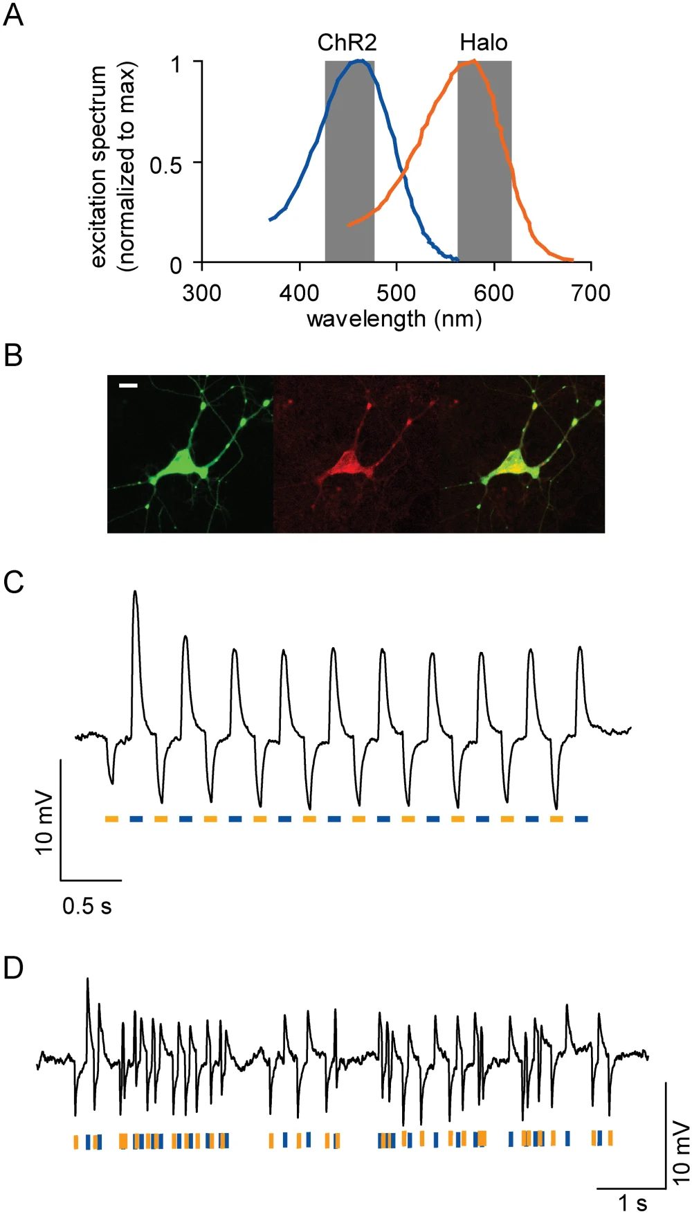
*Original paper figure: a single neuron expresses both the activating and silencing proteins. Panel A shows that blue light drives ChR2 and yellow light drives Halo, with absorption spectra that barely overlap, allowing independent control. Panel C shows the membrane potential rising and falling as blue and yellow light alternate. Source: Han X, Boyden ES. Multiple-Color Optical Activation, Silencing, and Desynchronization of Neural Activity, with Single-Spike Temporal Resolution. PLoS One. 2007;2(3):e299. DOI [10.1371/journal.pone.0000299](https://doi.org/10.1371/journal.pone.0000299); PMID 17375185; PMC1808431. CC BY 4.0. Reproduced at reduced size without modification.*
---
## Why Do Aged Mice Sleep Poorly?
That same population of arousal neurons reappeared in aging research fifteen years later. In 2022, de Lecea's lab published a study in *Science* whose opening sentence reads: sleep quality declines with age, but the mechanisms remain poorly understood. The team compared young mice aged 3 to 5 months with aged mice aged 18 to 22 months. The study can be read in four steps.
**Step one: what goes wrong with sleep in aged mice.** Their sleep is more fragmented; they wake up more often during sleep. The team recorded that hypocretin neurons in aged mice generated more frequent bursts of activity, each one pushing the animal into a bout of wakefulness.
**Step two: what optogenetics confirmed.** Optogenetically stimulating this cell population caused aged mice to stay awake longer. This is where optogenetics earns its place in the paper: it establishes that activating these cells is sufficient to prolong wakefulness.
**Step three: how the mechanism was found.** This step relied mainly on other methods. Electrophysiology and histology revealed that aging hypocretin neurons were hyperexcitable, and the cause pointed to a potassium channel: KCNQ2 levels were reduced, weakening the "M-current" carried by KCNQ2/3 channels. Two lines of validation followed. Disrupting *Kcnq2/3* genes selectively in these neurons of young mice produced the fragmented sleep pattern characteristic of aged mice. Going the other direction, giving aged mice flupirtine, a drug that opens KCNQ channels, reduced the excitability of this cell population. The paper's word for the restored sleep architecture was "rejuvenated."
**Step four: why this is not yet a usable treatment.** Flupirtine was originally an analgesic. The European Medicines Agency recommended withdrawal of its marketing authorization in 2018 because of serious liver injury risk, including liver failure. The study establishes a mechanism in mice. A mouse sleeping better is not evidence that this drug is appropriate for older people with poor sleep. Getting from here to a usable intervention requires finding a safe approach and validating it separately in humans.
---
## Alzheimer's Model Mice and Lost Memories: Not Stored, or Just Inaccessible?
In 2016, Susumu Tonegawa's lab published a study in *Nature* using transgenic mice modeling early Alzheimer's disease. These mice performed as though they had forgotten in a contextual fear memory test: returned to an environment where they had previously received a shock, they showed no expected fear response.
The team first used genetic labeling to tag the hippocampal cells that were active during learning (the "engram cells" holding that memory) with ChR2. Later, directly stimulating those cells with light produced the fear response. The conclusion: the problem in these mice was retrieval, not storage.
The team also found that this retrieval failure worsened progressively before amyloid plaques appeared, while dendritic spine density on engram cells declined over the same period. Optogenetically inducing long-term potentiation at synapses of those cells restored both spine density and long-term memory. Eliminating the rescued engram cells abolished the recovered memory.
This result is easy to read as "memories in Alzheimer's patients are actually still there." What the original data actually support is narrower: this was a mouse model carrying human Alzheimer's-associated gene mutations, studied at an early stage, for a specific memory type, and "calling memories back with light" required prior genetic labeling and an implanted optical fiber. The paper answers a mechanistic question. There is no equivalent procedure in humans.
---
## From Animal Research to Treatment: How Far Has the Field Come?
Based on ClinicalTrials.gov registrations and published literature as of October 9, 2026, human optogenetics trials are concentrated in retinal diseases. Searching ClinicalTrials.gov that day with terms including optogenetic, channelrhodopsin, ChrimsonR, and ChronosFP, I found more than a dozen registrations, all in the eye, none in the brain or ear. My reading of this: the eye is a natural first destination. Light already reaches it. The injection site is accessible. Visual acuity can be measured. The previous article in this series on [partial reprogramming's first human trial](/en/articles/partial-reprogramming-osk/) similarly targeted a single eye.
The earliest published human efficacy evidence is a single case reported in *Nature Medicine* in 2021. A patient blinded by retinitis pigmentosa received an intraocular injection of an adeno-associated virus carrying ChrimsonR, a red-light-responsive channelrhodopsin, and wore specialized light-emitting goggles. Using only the treated eye with the goggles on, the patient could detect, locate, count, and touch objects on a table.
Results from ten participants appeared two days after the Nobel announcement. On October 7, 2026 (US Eastern time), the *New England Journal of Medicine* published the PIONEER trial: 10 participants blinded by advanced retinitis pigmentosa each received a single injection into their worse-seeing eye. The trial registration remains in follow-up. The primary outcome was safety: 9 of 10 participants experienced a combined 34 ocular adverse events: 23 mild, 10 moderate, 1 serious. The serious event was a transient central retinal artery occlusion immediately after injection, resolving within minutes with medication. Among secondary outcomes, 7 of 10 participants showed increased light sensitivity, ranging from 2-fold to 62.3-fold. The threshold the authors used for "clinically meaningful" (roughly 4-fold) was not prespecified in the trial protocol; 6 participants met it. The conclusion: safe within the limitations of this small study; further research is needed to assess both safety and efficacy.
In his telephone interview on prize day, Nagel said he had been telling friends around him to stay calm, because "so far, as far as I know, there is only one patient who has benefited from optogenetics," unlike the large numbers helped by antimalarial drugs or immune checkpoint inhibitors, and he had therefore thought a medicine prize was still premature.
A second line of development is Nanoscope Therapeutics' MCO-010 (sonpiretigene isteparvovec, brand name MOGENRY), which delivers a light-sensitive protein gene to bipolar cells in the retina. In September 2026, Nanoscope announced that the FDA had accepted its biologics license application, with the indication being retinitis pigmentosa with severe vision loss in adults. Acceptance means the application has entered review, not that it has been approved.
The trials described so far address inherited retinal disease, not aging. The only trial in the registry directly relevant to aging began enrolling in January 2026: Bionic Sight's BS01 delivers a channelrhodopsin gene (ChronosFP) via adeno-associated virus into the eyes of patients aged 50 or older with geographic atrophy from dry age-related macular degeneration. This is a Phase 1/2 trial planning to enroll 40 participants; the second phase includes a sham-injection control group.
For the ear, optogenetic improvements to cochlear implants remain at the animal and engineering research stage. One registered study uses light to stimulate the auditory nerve in a cochlear implant (NCT05110183), but it involves no gene delivery and is a different technology, not optogenetics.
> **Key Data: Optogenetics Human Trials (as of October 9, 2026)**
> - **PIONEER** (*New England Journal of Medicine*, published October 7, 2026 US ET): 10 participants; 9 had 34 ocular adverse events total, including 1 serious but transient event; 7 showed increased light sensitivity (2.0 to 62.3-fold)
> - **MCO-010 (MOGENRY)**: Company announced FDA acceptance of BLA in September 2026; under review
> - **BS01** (geographic atrophy from dry AMD): Phase 1/2, began enrolling January 2026, 40 participants planned
> - **Brain, ear**: No human trials delivering light-sensitive genes found on ClinicalTrials.gov (search terms: optogenetic, channelrhodopsin, ChrimsonR, ChronosFP)
### For Brain Diseases, the Path Is Mostly Indirect
This does not mean optogenetics is irrelevant to brain disease. It just helps in an indirect way. In late 2025, eleven researchers including Deisseroth published a roadmap in *Nature Neuroscience*. The second sentence of the abstract states: many translation pathways do not depend on applying optogenetics directly in humans, but rather on using causal knowledge gained through optogenetics to develop and advance other therapeutic approaches.
Parkinson's disease is one example. Deep brain stimulation has been used in patients for years, but what types of cells an electrode placed in the subthalamic nucleus actually stimulates has been difficult to determine. In 2009, Deisseroth's team used optogenetics in a Parkinson's model mouse to turn different cell types and fiber pathways in that region on and off one by one. They found that the therapeutic effect could be explained by stimulating the afferent axons projecting into that nucleus, not the local cells. In 2010, Anatol Kreitzer's lab and Deisseroth together used optogenetics to demonstrate that the "direct pathway" and "indirect pathway" of the basal ganglia have opposing effects on movement: activating the indirect pathway produced Parkinsonian freezing and bradykinesia; activating the direct pathway fully rescued those deficits in Parkinson's model mice.
The sleep study follows the same logic. Optogenetics helped identify which cells are driving wakefulness; addressing that requires a drug or other intervention. And the drug used in that study carries its own safety problems.
---
## How Is 40 Hz Light and Sound Stimulation Related to Optogenetics?
This research line started with an optogenetics experiment and later switched to light and sound as sensory stimuli, without delivering any light-sensitive gene into neurons. The human trials therefore evaluate a different intervention.
In 2016, Li-Huei Tsai's lab at MIT published a study in *Nature* in two parts. The first used optogenetics to drive a population of hippocampal interneurons in 5XFAD Alzheimer's model mice at 40 pulses per second; amyloid-beta in the brain decreased, and no other frequency produced this effect. The second part had mice view light flashing 40 times per second. At this point the approach changed. Optogenetics had installed a light-gated channel in specific neurons and opened them with an optical fiber. The flashing light instead enters through the eyes as visual stimulation, in the hope of entraining brain rhythms, with no light-gated channel in the neurons. This visual stimulation reduced amyloid in the visual cortex and lessened plaque burden in mice that already had plaques.
The subsequent mouse evidence has been inconsistent. In 2023, György Buzsáki's lab at New York University replicated the experiment in two model strains. In visual cortex, entorhinal cortex, and hippocampus, the 40 Hz flashing light neither entrained endogenous gamma rhythms nor produced reliable changes in plaque count, microglial morphology, or amyloid levels. The mice also avoided the 40 Hz flashing light. In 2024, Tsai's team proposed another mechanism in *Nature*: combined 40 Hz light and sound stimulation promotes influx of cerebrospinal fluid and outflow of interstitial fluid (that is, glymphatic clearance), and blocking this clearance pathway eliminated the amyloid-removal effect. The conflict of interest statement in that paper discloses that Tsai and Boyden are co-scientific founders of Cognito Therapeutics.
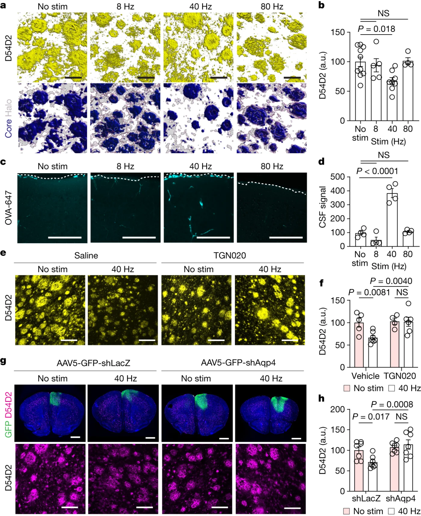
*Original paper figure: panels a–d show 5XFAD model mice exposed to 8, 40, and 80 Hz combined light and sound; only 40 Hz reduced amyloid plaques and elevated CSF signal. Panels e–h show that pharmacological or genetic suppression of the water channel AQP4 abolished the 40 Hz effect. This is a mouse experiment. The authors disclose a financial relationship with Cognito Therapeutics. Source: Murdock MH, et al. Multisensory gamma stimulation promotes glymphatic clearance of amyloid. Nature. 2024;627(8002):149–156. DOI [10.1038/s41586-024-07132-6](https://doi.org/10.1038/s41586-024-07132-6); PMID 38418876; PMC10917684. CC BY 4.0. Reproduced at reduced size without modification.*
Cognito has developed 40 Hz light and sound stimulation into a home-use device. Human evidence currently has two layers.
**Primary outcome.** The small feasibility trial OVERTURE enrolled 76 patients with mild-to-moderate Alzheimer's disease over 6 months. On the primary clinical outcome, there was no statistically significant difference between the treatment and sham device groups. The paper also notes that the trial was not powered to evaluate efficacy.
**Exploratory results.** Several secondary outcomes, including daily functioning, the Mini-Mental State Examination, and total brain volume, reached "nominal" significance, meaning the p-values were not corrected for multiple comparisons. Results of this kind support hypothesis generation; they are not evidence of efficacy.
**The missing piece.** The trial designed to actually answer whether the treatment works is HOPE: randomized, double-blind, sham-controlled, 673 participants, using the Spectris AD device at home for approximately 12 months per day. The trial completed in August 2026. As of October 9, no results have been posted to the registry, and I found no company announcement of primary results. The company website still describes it as "an investigational therapy."
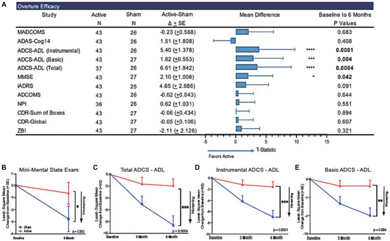
*Original paper figure: the first row shows the primary outcome, where the two groups show no difference. The rows below, with smaller p-values, are secondary outcomes without correction for multiple comparisons; panels B through E plot their trajectories over time. This is a small trial not designed to assess efficacy, funded by Cognito Therapeutics, with most authors being company employees. Source: Hajós M, et al. Safety, tolerability, and efficacy estimate of evoked gamma oscillation in mild to moderate Alzheimer's disease. Front Neurol. 2024;15:1343588. DOI [10.3389/fneur.2024.1343588](https://doi.org/10.3389/fneur.2024.1343588); PMID 38515445; PMC10957179. CC BY. Reproduced at reduced size without modification.*
> **Key Data: The 40 Hz Research Line (as of October 9, 2026)**
> - **OVERTURE** (published 2024): 76 participants, 6 months. Primary outcome MADCOMS showed no difference (p=0.683); secondary outcomes CDR-SB and ADAS-Cog14 showed no difference; ADCS-ADL, MMSE, and total brain volume reached nominal significance without multiple comparison correction
> - **HOPE** ([NCT05637801](https://clinicaltrials.gov/study/NCT05637801)): randomized, double-blind, sham-controlled; 673 participants; daily use of Spectris AD device for 60 minutes, approximately 12 months; primary endpoint iADMARS (a composite of MMSE and daily functioning); completed August 17, 2026; registry last updated September 9; no results posted
> - **Mouse studies**: 2016 (Tsai lab, 5XFAD, reduction in Aβ1-40 and Aβ1-42), 2023 (Buzsáki lab, APP/PS1 and 5xFAD, no reliable entrainment or Aβ40/42 change), 2024 (Tsai lab, 5XFAD, glymphatic clearance)The current state of evidence: mouse results point in both directions, the small human trial showed no difference on its primary clinical outcome, and results from the large trial are not yet available. Based on the evidence laid out in this article, claiming that 40 Hz stimulation can already improve dementia goes beyond what the evidence supports. Equally, HOPE's data have not been released, so the treatment cannot be declared ineffective on current information either.
---
## How to Read This Year's Prize and Related Therapeutic Claims
This Nobel Prize recognizes a research tool and the causal experimental capacity it enables. It has answered specific questions in mice and, in humans, has reached early trials in retinal disease. The prize itself says nothing about whether any therapy invoking "light" is effective.
After optogenetics wins a Nobel, I expect the word "light" to be borrowed for other things: 40 Hz flashing light, red light, near-infrared irradiation. Whether any of these work depends on their own evidence. When I encounter claims of this kind, I ask three questions:
1. **Was a light-sensitive protein gene delivered into cells?** If not, it is not optogenetics.
2. **Where does the light go, and how does it get there?** An optical fiber inserted into an animal brain, or goggles projecting light onto a genetically treated retina, is a different thing from illuminating skin or scalp from outside, or having the eyes watch a flashing stimulus.
3. **Where does the evidence stop?** Mice, a single case, a small trial, a large randomized controlled trial, regulatory approval: each step up the ladder requires fresh proof.
This is the same evidence hierarchy as the first article in this series on the [hallmarks of aging](/en/articles/hallmarks-of-aging-map/): the higher you go, the closer the evidence gets to "people are actually healthier."

*Figure 2: Four approaches that each involve light. Each column addresses one of the three questions: whether a gene is delivered, how the light reaches its target, and where the evidence currently stands. Data as of October 9, 2026; sources in the references below.*
---
## What Has Optogenetics Given Aging Research?
Optogenetics has given researchers a better way to identify which cells are involved in sleep and memory problems. In aged mice, it helped point to a population of neurons that makes animals too easy to wake. In early Alzheimer's model mice, it showed that lost memories may still be stored, only unretrievable.
Turning that knowledge into safe, effective human treatment still requires more research: finding interventions that can be used in people, establishing their safety, and demonstrating efficacy in trials large enough to give a reliable answer.
This is the fifth article in the "[Molecules and Aging](/series/molecular-aging/)" series, filed under my [contemplation and memory](/en/topics/life/) topic. The second article in the series covers plasma proteins as predictors of mortality and is collected in [Predictive Power and Causal Inference Are Two Different Things](/en/articles/plasma-omics-aging-mortality/). Optogenetics addresses exactly the second of those two things.
---
## Supplementary Material
### Molecular Details of Channelrhodopsin
The rhodopsin in our eyes and the channelrhodopsin in green algae share a similar architecture: both are proteins that cross the cell membrane seven times, and both hold a retinal molecule in the middle of the seventh transmembrane helix, attached through a Schiff base linkage to a lysine residue. Whether the two share a common ancestor is a question the 2014 review by Ernst et al. found the available data insufficient to settle.
The differences come after that. Animal rhodopsin holds 11-cis retinal; light converts it to all-trans, triggering a G-protein signaling cascade: rhodopsin activates the G-protein, the G-protein activates an enzyme, the enzyme changes the concentration of a second messenger, and only then does an ion channel change state. Microbial rhodopsins hold all-trans retinal; light converts it to 13-cis. In the channelrhodopsin subtype, the protein is itself the channel: the conformational change opens the pore directly.
Nagel's 2002 paper reported that ChR1 is primarily a proton channel. The 2003 paper reported that ChR2 conducts both monovalent and divalent cations; the final sentence of the abstract states that ChR2 can depolarize cells of any size by light alone. In 2015, researchers found a naturally occurring light-gated anion channel in another alga, adding a second option for the silencing toolkit.
### Prize Background
- **Laureates and prize sum:** Deisseroth is affiliated with the Howard Hughes Medical Institute (HHMI) and Stanford University. Hegemann's early *Chlamydomonas* work was conducted at the Max Planck Institute of Biochemistry in Martinsried near Munich; Nagel was at the Max Planck Institute of Biophysics in Frankfurt at the time. The prize sum is 12 million Swedish kronor, divided equally among the three. Committee chair Per Svenningsson stated in the press release that optogenetics lets scientists map the brain in ways that were previously only a dream.
- **Recollections from prize day:** Nagel said in his telephone interview that it was difficult at first, and then it was "lucky: I used the wrong, somewhat acidic solutions, and suddenly there was a clear signal." Hegemann recalled that the journal *Science* initially described the finding as "ion channels from pond scum." The Nobel Committee's popular science document notes that Deisseroth's starting point was his psychiatric training, where he saw patients' suffering and the limited effectiveness of available treatments.
- **Contributors outside the prize:** The Nobel Prize in each category can be awarded to at most three people. Edward Boyden, now at MIT, was first author on the landmark 2005 paper. Ernst Bamberg, Nagel's doctoral supervisor, was a co-author on all three key papers from 2002, 2003, and 2005. Gero Miesenböck demonstrated as early as 2002 that genetic engineering could make normally non-photosensitive neurons respond to light, though his system required three genes working together and responded more slowly. The Brain Prize in 2013, the first major award for optogenetics, went to six scientists simultaneously, including all three of this year's Nobel laureates.
- **Visit to Taiwan:** Taiwan's Central News Agency reported that Deisseroth is expected to visit Taiwan in March 2027 to give a lecture in Academia Sinica's Chemical Biology series.
### Further Reading: A Lifelong Behavioral Study in African Killifish
In March 2026, labs led by Deisseroth and aging biologist Anne Brunet published a study in *Science* tracking the behavior of African turquoise killifish from adolescence to death. A machine learning model trained only on behavior recorded early in life could predict each individual fish's lifespan. This is a predictive study; it does not use optogenetics to manipulate cells. It belongs to the same class of question as the plasma proteomics work in the second article of this series.
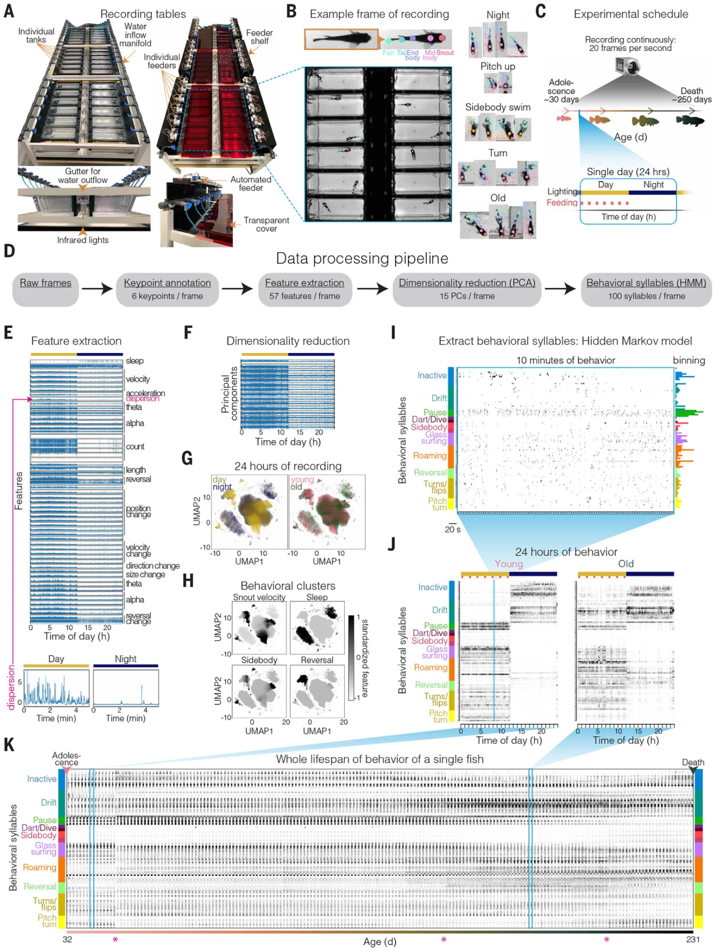
*Original paper figure: read from left to right to follow how the data were generated. Each fish was video-recorded around the clock from adolescence to death. The footage was then decomposed into hundreds of recurring "behavioral syllables." Panel K shows the lifetime behavioral record of a single fish. This is a behavioral recording and prediction study, not an optogenetics experiment. Source: Bedbrook CN, ..., Brunet A, Deisseroth K. Lifelong behavioral screen reveals an architecture of vertebrate aging. Science. 2026;391(6790):eaea9795. DOI [10.1126/science.aea9795](https://doi.org/10.1126/science.aea9795); PMID 41818367; PMC13165398 (accepted manuscript). CC BY 4.0. Reproduced at reduced size without modification.*
---
## References
**Official Nobel Documents**
- The Nobel Assembly at Karolinska Institutet. Press release: The Nobel Prize in Physiology or Medicine 2026. 2026-10-05.
- The Nobel Committee for Physiology or Medicine. Popular information: A light-sensitive algal protein energised neuroscience. 2026. (source for the ~0.5 ms response in *Chlamydomonas*, ≥10 ms in the human eye, and ~0.2 ms for ChR2)
- The Nobel Committee for Physiology or Medicine. Scientific background 2026: Optogenetics. Discovery of a neuronal switch. (Crick's vision, endogenous retinal, the methodological lineage from "reading" to "writing," contributors outside the prize)
- Nobel Prize first reactions (telephone interview transcripts): [Karl Deisseroth](https://www.nobelprize.org/prizes/medicine/2026/deisseroth/interview/), [Peter Hegemann](https://www.nobelprize.org/prizes/medicine/2026/hegemann/interview/), [Georg Nagel](https://www.nobelprize.org/prizes/medicine/2026/nagel/interview/)
**Origins of Channelrhodopsin and Optogenetics**
- Nagel G, Ollig D, Fuhrmann M, Kateriya S, Musti AM, Bamberg E, Hegemann P. Channelrhodopsin-1: a light-gated proton channel in green algae. *Science*. 2002;296(5577):2395–2398. PMID 12089443.
- Nagel G, Szellas T, Huhn W, Kateriya S, Adeishvili N, Berthold P, Ollig D, Hegemann P, Bamberg E. Channelrhodopsin-2, a directly light-gated cation-selective membrane channel. *Proc Natl Acad Sci U S A*. 2003;100(24):13940–13945. PMID 14615590.
- Boyden ES, Zhang F, Bamberg E, Nagel G, Deisseroth K. Millisecond-timescale, genetically targeted optical control of neural activity. *Nat Neurosci*. 2005;8(9):1263–1268. PMID 16116447.
- Deisseroth K, Feng G, Majewska AK, Miesenböck G, Ting A, Schnitzer MJ. Next-generation optical technologies for illuminating genetically targeted brain circuits. *J Neurosci*. 2006;26(41):10380–10386. PMID 17035522. (first formal use of the term "optogenetics")
- Aravanis AM, et al. An optical neural interface: in vivo control of rodent motor cortex with integrated fiberoptic and optogenetic technology. *J Neural Eng*. 2007;4(3):S143–S156. PMID 17873414.
- Adamantidis AR, Zhang F, Aravanis AM, Deisseroth K, de Lecea L. Neural substrates of awakening probed with optogenetic control of hypocretin neurons. *Nature*. 2007;450(7168):420–424. PMID 17943086.
- Zhang F, et al. Multimodal fast optical interrogation of neural circuitry. *Nature*. 2007;446(7136):633–639. PMID 17410168. (halorhodopsin, Deisseroth lab)
- Han X, Boyden ES. Multiple-color optical activation, silencing, and desynchronization of neural activity, with single-spike temporal resolution. *PLoS One*. 2007;2(3):e299. PMID 17375185. (halorhodopsin, Boyden lab)
- Govorunova EG, Sineshchekov OA, Janz R, Liu X, Spudich JL. Natural light-gated anion channels: a family of microbial rhodopsins for advanced optogenetics. *Science*. 2015;349(6248):647–650. PMID 26113638. (naturally occurring light-gated anion channels)
- Zemelman BV, Lee GA, Ng M, Miesenböck G. Selective photostimulation of genetically chARGed neurons. *Neuron*. 2002;33(1):15–22. PMID 11779476.
- Ernst OP, et al. Microbial and animal rhodopsins: structures, functions, and molecular mechanisms. *Chem Rev*. 2014;114(1):126–163. PMID 24364740. (11-cis and all-trans retinal, Schiff base and lysine; review)
**Aging and Neurodegeneration Research (all animal studies)**
- Li SB, Damonte VM, et al., de Lecea L. Hyperexcitable arousal circuits drive sleep instability during aging. *Science*. 2022;375(6583):eabh3021. PMID 35201886.
- Roy DS, Arons A, Mitchell TI, Pignatelli M, Ryan TJ, Tonegawa S. Memory retrieval by activating engram cells in mouse models of early Alzheimer's disease. *Nature*. 2016;531(7595):508–512. PMID 26982728.
- Iaccarino HF, et al., Tsai LH. Gamma frequency entrainment attenuates amyloid load and modifies microglia. *Nature*. 2016;540(7632):230–235. PMID 27929004. (two author corrections: a 2018 correction to Extended Data figures with restated statistics, authors reported conclusions unchanged; a 2024 author name update)
- Soula M, et al., Buzsáki G. Forty-hertz light stimulation does not entrain native gamma oscillations in Alzheimer's disease model mice. *Nat Neurosci*. 2023;26(4):570–578. PMID 36879142.
- Murdock MH, et al., Tsai LH. Multisensory gamma stimulation promotes glymphatic clearance of amyloid. *Nature*. 2024;627(8002):149–156. PMID 38418876. (conflict of interest: Tsai and Boyden are co-scientific founders of Cognito Therapeutics)
- Gradinaru V, Mogri M, Thompson KR, Henderson JM, Deisseroth K. Optical deconstruction of parkinsonian neural circuitry. *Science*. 2009;324(5925):354–359. PMID 19299587.
- Kravitz AV, et al., Kreitzer AC. Regulation of parkinsonian motor behaviours by optogenetic control of basal ganglia circuitry. *Nature*. 2010;466(7306):622–626. PMID 20613723.
- Bedbrook CN, Nath RD, Zhang L, Linderman SW, Brunet A, Deisseroth K. Lifelong behavioral screen reveals an architecture of vertebrate aging. *Science*. 2026;391(6790):eaea9795. PMID 41818367. (African turquoise killifish; predictive study, further reading)
**Human Trials and Translation**
- Sahel JA, et al., Roska B. Partial recovery of visual function in a blind patient after optogenetic therapy. *Nat Med*. 2021;27(7):1223–1229. PMID 34031601. (single case)
- Sahel JA, et al., Roska B. Optogenetic therapy for restoring aspects of visual function. *N Engl J Med*. 2026;395(14):1399–1408. PMID 42842903. (PIONEER, NCT03326336; open-label, 10 participants; funded by GenSight Biologics; published online October 7, 2026 US ET)
- University of Pittsburgh (EurekAlert! press release). [Optogenetic therapy safe, shows signs of vision restoration in patients with advanced retinitis pigmentosa, according to international study led by Pitt scientists](https://www.eurekalert.org/news-releases/1146208). 2026-10-07
- Hajós M, et al. Safety, tolerability, and efficacy estimate of evoked gamma oscillation in mild to moderate Alzheimer's disease. *Front Neurol*. 2024;15:1343588. PMID 38515445. (OVERTURE; most authors are Cognito employees)
- Lüscher C, et al., Deisseroth K. Roadmap for direct and indirect translation of optogenetics into discoveries and therapies for humans. *Nat Neurosci*. 2025;28(12):2415–2431. PMID 41254285.
- ClinicalTrials.gov registrations: [NCT05637801](https://clinicaltrials.gov/study/NCT05637801) (HOPE), [NCT07158775](https://clinicaltrials.gov/study/NCT07158775) (BS01, geographic atrophy), [NCT03326336](https://clinicaltrials.gov/study/NCT03326336) (PIONEER), [NCT04945772](https://clinicaltrials.gov/study/NCT04945772) (MCO-010 RESTORE trial), [NCT05110183](https://clinicaltrials.gov/study/NCT05110183) (optical cochlear implant, no gene delivery). Accessed 2026-10-09.
- Ophthalmology Times. FDA accepts BLA for MCO-010 in retinitis pigmentosa. 2026-09-09.
- European Medicines Agency. Flupirtine-containing medicinal products (2018 recommendation for withdrawal of marketing authorization).
- Cognito Therapeutics. Clinical studies. (accessed 2026-10-09)
- 1111 產經新聞網. [MIT學者、中興大學校友蔡立慧 發現阿茲海默症逆轉新法](https://www.1111.com.tw/news/jobns/150987). 2023-04-21 (Li-Huei Tsai's academic and professional background)
**International and Domestic News Coverage**
- Central News Agency. [三位美德學者獲諾貝爾醫學獎 光遺傳學研究為腦科學開新局](https://www.cna.com.tw/news/aopl/202610050264.aspx). 2026-10-05
- Central News Agency. [諾貝爾醫學獎得主戴瑟羅斯低調如「角落生物」明年訪台傳授光遺傳](https://www.cna.com.tw/news/ahel/202610050299.aspx). 2026-10-05 (source for the Taiwan visit)
- PTS News Network. [開拓光遺傳學為腦疾帶來曙光 美、德3學者獲諾貝爾生醫獎](https://news.pts.org.tw/article/830061). 2026-10-06
- UDN. [光遺傳學奪諾貝爾醫學獎 國內專家:有望助攻失智、憂鬱與失明治療](https://udn.com/news/story/6656/9796451). 2026-10-05
- The Associated Press (via WUNC). [Nobel medicine prize goes to 3 scientists for research into brain activity](https://www.wunc.org/2026-10-05/nobel-medicine-prize-goes-to-3-scientists-for-research-into-brain-activity). 2026-10-05
- Reuters (via Khaleej Times). [German, US scientists win Nobel medicine prize for work on optogenetics](https://www.khaleejtimes.com/world/german-us-scientists-win-nobel-medicine-prize-optogenetics). 2026-10-05
- BBC News, Michelle Roberts. [Nobel Prize for Medicine for showing inner workings of the brain](https://www.bbc.co.uk/news/articles/c5ev3ypmzly8o). 2026-10-05
- The Guardian. [Nobel prize in medicine 2026 awarded for research into mysteries of brain](https://www.theguardian.com/science/2026/oct/05/nobel-prize-medicine-2026-winner). 2026-10-05
- Al Jazeera. [Nobel medicine prize honours US and German scientists for optogenetics work](https://www.aljazeera.com/news/2026/10/5/nobel-medicine-prize-honours-us-and-german-scientists-for-optogenetics-work). 2026-10-05
- DW, Jenipher Camino Gonzalez. [German and US scientists win Nobel Prize 2026 in medicine](https://www.dw.com/en/german-and-us-scientists-win-nobel-prize-in-medicine-for-research-on-brain-activity/a-79544130). 2026-10-05
- Nature News, Miryam Naddaf & Ewen Callaway. [Medicine Nobel awarded for brain 'switch' that controls neurons with light](https://www.nature.com/articles/d41586-026-03091-2). 2026-10-05
- Nature, Ewen Callaway. ['What took them so long?' This year's medicine Nobel winner is overdue](https://www.nature.com/articles/d41586-026-03094-z). 2026-10-05 (source for the six Brain Prize laureates in 2013)
- STAT, Annalisa Merelli. [2026 Nobel Prize in Medicine awarded for brain research tool called optogenetics](https://www.statnews.com/2026/10/05/nobel-prize-medicine-2026-winner-deisseroth-hegemann-nagel/). 2026-10-05
- Scientific American, Jackie Flynn Mogensen. [2026 Nobel Prize in Physiology or Medicine awarded to scientists behind optogenetics](https://www.scientificamerican.com/article/2026-nobel-prize-in-physiology-or-medicine-awarded-to-karl-deisseroth-george-nagel-and-peter-hegemann-for-work-on-optogenetics/). 2026-10-05
- The Scientist. [Nobel Prize for Optogenetics](https://www.the-scientist.com/nobel-prize-for-optogenetics-75099). 2026-10-05
- Stanford Report, Krista Conger. [Karl Deisseroth awarded 2026 Nobel Prize in physiology or medicine](https://news.stanford.edu/stories/2026/10/karl-deisseroth-nobel-prize-physiology-medicine). 2026-10-05 (source for "precise causal perturbation" and "we are poking the brain")
- HHMI. [Karl Deisseroth Awarded the 2026 Nobel Prize in Physiology or Medicine](https://www.hhmi.org/news/karl-deisseroth-2026-nobel-prize-physiology-medicine)
- Max Planck Society. [Congratulations to Peter Hegemann and Georg Nagel on the 2026 Nobel Prize in Physiology or Medicine](https://www.mpg.de/27014852/congratulations-to-peter-hegemann-and-georg-nagel-on-their-nobel-prize-in-physiology-or-medicine)
- Universität Würzburg. [Nobelpreis für den Würzburger Professor Georg Nagel](https://www.uni-wuerzburg.de/aktuelles/pressemitteilungen/single/news/nobelpreis-georg-nagel/). 2026-10-05
**Videos**
Official announcement and laureate interviews
- Nobel Prize (YouTube): [Announcement of the 2026 Nobel Prize in Physiology or Medicine](https://www.youtube.com/watch?v=qGPpW0hAaaY). 2026-10-05, approx. 30 min. Full livestream of the announcement.
- Nobel Prize (YouTube): ["We can get a whole new understanding of the function of the brain." Medicine prize 2026](https://www.youtube.com/watch?v=Q3yoss9R8F4). 2026-10-05, approx. 5 min. Committee member explains the switch.
- Nobel Prize (YouTube): First telephone interviews with the three laureates: [Karl Deisseroth](https://www.youtube.com/watch?v=RNogbM246cE), [Peter Hegemann](https://www.youtube.com/watch?v=bZ13m69ZwCk), [Georg Nagel](https://www.youtube.com/watch?v=E_tL0QzwOZU). 2026-10-05, approx. 4 to 6 min each.
- Stanford (YouTube): [Karl Deisseroth on winning the 2026 Nobel Prize in physiology or medicine](https://www.youtube.com/watch?v=LiiKmHpoQjE). 2026-10-05, approx. 3 min.
Optogenetics introductions
- nature video: [Method of the Year 2010: Optogenetics](https://www.youtube.com/watch?v=I64X7vHSHOE). 2010, approx. 4.5 min. Animated explainer.
- iBiology Science Stories: [Karl Deisseroth (Stanford / HHMI): Development of Optogenetics](https://www.youtube.com/watch?v=MUGky_QaaV0). 2016, approx. 21 min. The laureate describes how the method came together.
- NeuroCure: [Optogenetics - Visit the Lab with Peter Hegemann](https://www.youtube.com/watch?v=2DXn9zIPL6Q). 2023, approx. 4 min. A walk through Hegemann's laboratory.
- TED-Ed: [A light switch for neurons - Ed Boyden](https://www.youtube.com/watch?v=az9tHuOzOfA). 2013, approx. 18 min. A talk by the first author of the 2005 paper.
Aging and neurodegeneration
- IOB Institute of Ophthalmology Basel: [Optogenetic vision restoration explained - Botond Roska](https://www.youtube.com/watch?v=uEAszLzpjec). 2021, approx. 4 min. Overview of the 2021 single-case study.
- TED: [Could We Treat Alzheimer's with Light and Sound? | Li-Huei Tsai](https://www.youtube.com/watch?v=AS0K0XOMNwA). 2022, approx. 12.5 min. The researcher's own talk, recorded before the 2023 contradicting study and before the HOPE trial completed.
- Falling Walls Foundation: [Tobias Moser: Breaking the Wall of Hearing Restoration](https://www.youtube.com/watch?v=qY0flgRjImY). 2022, approx. 3 min. The concept of an optogenetic cochlear implant; still at the preclinical stage.